Other news
Article in Advanced Materials
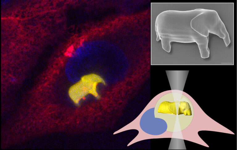
Researchers Maruša Mur, Aljaž Kavčič, Uroš Jagodič, Rok Podlipec and Matjaž Humar from Condensed Matter Physics Department of the Jožef Stefan Institute have shown that 3D printing can be performed inside a living human cell. First, they injected droplets of a bio-compatible photo-curable material into cells. Then, using a highly focused laser beam, they selectively illuminated the printing material and polymerized it. By moving the laser beam in three dimensions, it is possible to "draw" complex structures of any shape with sub-micrometer resolution. Using this method, the team printed various structures, from geometric patterns to microlasers and even small elephants, all inside living human cells. The cells containing such structures can migrate and undergo cell division where the structure is passed into one of the daughter cells. By transforming living cells into miniature environments for 3D printing, this work pushes the boundaries of what is possible at the intersection of biology, physics, and engineering, offering a powerful new tool for exploring the workings of life from the inside out. The results were published in a paper in Advanced Materials, that was selected as an Editor’s Choice Paper.
The article was published in the journal Advanced Materials. M. Mur, A. Kavčič, U. Jagodič, R. Podlipec, and M. Humar, “ Two-Photon 3D Printing of Functional Microstructures Inside Living Cells.” Advanced Materials (2026): e19286. https://doi.org/10.1002/adma.202519286
Biophotonics of lipid droplets: from natural optical resonators to highly precise sensors of dynamic processes in adipocytes
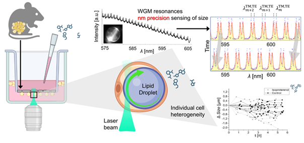
Colleagues from the department, Rok Podlipec, Ana Krišelj, and Matjaž Humar, in collaboration with the Department of Biochemistry, Molecular and Structural Biology and colleagues from the Helmholtz Center in Munich, have published an article in ACS Sensors on an exceptionally precise method for studying rapid dynamic processes at the level of individual adipocytes. In the study, they employed laser‑excited so‑called “whispering gallery mode” (WGM) optical resonances in lipid droplets of live primary adipocytes, achieving nanometer‑scale accuracy in measuring droplet size, which significantly surpasses the resolution of conventional microscopy. By monitoring their dynamics, they explored complex responses to pharmacological agents, variability among individual cells—undetectable with standard bulk assays—and early changes in cell viability, faster than conventional tests. The presented method paves the way for investigations of metabolism and obesity‑related diseases at the level of single adipocytes and tissues.
More information can be found here: https://pubs.acs.org/doi/10.1021/acssensors.5c03272